


Here's the text.
MR testing
Last Updated June 18, 2020
Overview of Inspection
The image obtained by MR examination is called MRI, but MRI is an abbreviation of Magnetic Resonance Imaging and is translated as a magnetic resonance image in Japanese.
An MR test is a test that uses the power of magnetic and electromagnetic waves to obtain a cross-sectional image inside the body.
Since X-rays are not used, there is no radiation exposure.
Head MR test
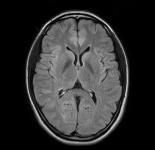 The image contrast is excellent, and it is possible to draw a detailed structure of the brain.
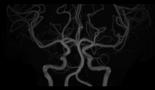 It is possible to draw the form of blood vessels without using a contrast agent.
Lumbar vertebral MR test
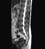 The image contrast is excellent, and it is possible to draw discs and nerves in addition to bone.
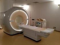
Facilities of our hospital
In our hospital, we have introduced two MR devices with different static magnetic field strength.
We strive to be able to conduct tests that take advantage of the characteristics of each device according to the location to be taken, the purpose of the test, and the symptoms of the patient.
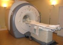
SIGNA ( Explorer
Made by GE Healthcare Japan
- 1.5 This is an MR device of T
- It adopts a detachable sleeper to reduce the burden on patients who have difficulty moving.
- In addition, since the sleeper goes down to a maximum of 49 cm, it is easy for even small children to get on and off.
- Equipped with a large number of advanced body motion correction technologies, it can handle a variety of patients and tests.
Ingenia 3.0T
Made by Phillips
- This is a 3.0T ( MR device.
- The high magnetic field can provide a high resolution image.
- Inclined magnetic field noise perceived by patients is reduced more than before.
- Since the opening diameter is 70cm, the pressure during inspection is eased.
Inquiries to this page
Stroke and Neurospinal Center
Telephone: 045-753-2500 (Representative)
Telephone: 045-753-2500 (Representative)
Fax: 045-753-2894
Page ID: 821-390-666


