


Here's the text.
CT examination
Last update date April 1, 2024
Overview of Inspection
CT is an abbreviation for Computed Tomography and is translated as computer tomography in Japanese.
By irradiating X-rays from around the human body and imaging the internal structure of the human body from the strength of the transmitted X-rays, it is possible to observe the state inside the body in a cross section of all angles.
Head CT examination
It is possible to know the state in the brain by taking pictures of more than a dozen seconds.
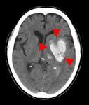
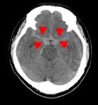
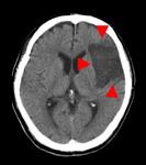
In the image example, it can be seen that intracerebral hemorrhage and subarachnoid hemorrhage are occurring because there is a white part of the normal part of the brain.
On the other hand, it can be seen that there is a cerebral infarction because there is a part drawn in black.
By adjusting the shade of the image, you can also check for skull fractures.
In addition, by injecting a contrast agent, more detailed observation of arteries and tumors is possible.
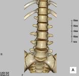
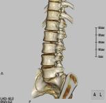
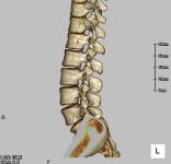
Spinal CT examination
Transformation and fracture of the vertebra, disordered arrangement, tumors, etc. can be observed in any cross section.
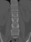
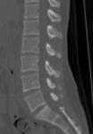
By image processing, it is also possible to display it three-dimensionally.
Facilities of our hospital
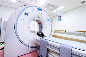
Aquilion ONE wan)
Made by Canon Medical Systems
This is a 320-row CT introduced in March 2021.
Since there is no sleeper movement and a wide area of 160 mm can be photographed in a single rotation, clear images can be obtained even in a short time. In particular, it is effective for emergency tests, blood flow evaluation of the entire brain using contrast agents, and evaluation of the heart.
In addition, the image reconstruction process for reducing exposure has been greatly improved, and the trunk can be photographed with a quarter of the conventional radiation dose.
In addition, it is now possible to perform more advanced tests in orthopedic areas, such as fracture evaluation in dual-energy photography and dynamic evaluation of bone joints by 4D photography.
Inquiries to this page
Stroke and Neurospinal Center
Telephone: 045-753-2500 (Representative)
Telephone: 045-753-2500 (Representative)
Fax: 045-753-2894
Page ID: 924-180-111


