


Here's the text.
Vascular imaging tests
Last update date April 1, 2024
Overview of Inspection
By inserting a soft and thin tube called a catheter from the blood vessels at the base of the arms and legs, proceeding with the catheter to the target blood vessel while looking at the X-ray fluoroscopy image, and injecting a contrast agent from the tip of the catheter to shoot, It is a test to see the form of blood vessels from various directions.
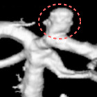
You can also display the blood vessels three-dimensionally to observe the complex shape of aneurysm and narrowed blood vessels in detail.
In addition to contrast agents, equipment such as coils, stents, and balloons can be inserted through the catheter, so it is also possible to treat intravascular diseases for abnormalities found during vascular photography.
In our hospital, we provide intravascular treatment such as coil embolization for cerebral aneurysm, stent placement for carotid artery stenosis, and thrombosis recovery therapy for acute cerebral infarction.
Cerebral aneurysmembolization
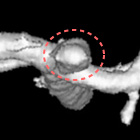
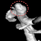
In order to prevent cerebral aneurysm from rupture and bleeding, this is an operation to pack coils into the cerebral aneurysm to prevent blood from entering.
When vascular photography is performed after surgery, the cerebral aneurysm replaced by coils will not be drawn because there is no contrast agent.
Carotid artery stent placement
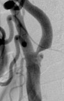 Preoperative
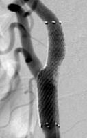 After surgery
This is an operation to restore blood flow by placing a metallic mesh tube called a stent in a carotid artery that has become narrower and poor blood flow, expanding blood vessels.
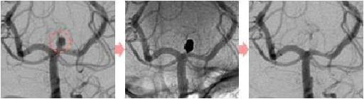
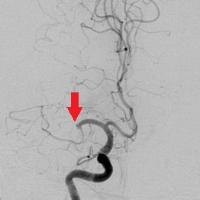
Thrombos recovery therapy
 Preoperative
 After surgery
When blood clots are clogged in the blood vessels of the brain and blood flow is interrupted, brain cells rapidly necrosis and cause cerebral infarction.
This treatment aims to remove clogged blood clots as soon as possible to resume blood flow and minimize damage to the brain so that serious sequelae from cerebral infarction can be avoided as much as possible.
Proceed the catheter to the blocked blood vessels and use equipment to scrape or suck out the blood clots to remove them.
BKP
The laboratory that performs vascular imaging tests has the same cleanliness as the operating room.
Therefore, in addition to vascular photography, some surgery in the Orthopaedic surgery area may be performed.
BKP stands for Balloon Kyphoplasty, and in Japanese it is called percutaneous vertebra formation.
This is an operation in which the vertebrae compression fracture due to osteoporosis is packed with bone cement while checking with X-ray fluoroscopy images, and approaches the shape of the vertebra before the fracture.
By stabilizing the vertebra, it can be expected to relieve pain.
Facilities of our hospital
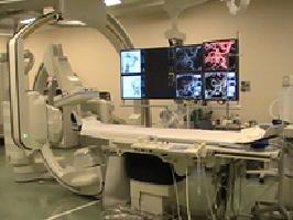
Artis zee biplane Artis)
Made by Siemens
- This device is equipped with an HDR flat panel detector, which has four times the gradient of shades of shades and can provide images with excellent contrast resolution.
- In addition, it has multiple automatic emission control mechanisms, which can optimize radiation dose for inspection.
Inquiries to this page
Stroke and Neurospinal Center
Telephone: 045-753-2500 (Representative)
Telephone: 045-753-2500 (Representative)
Fax: 045-753-2894
Page ID: 452-698-626


